Understanding the Human Skeleton: A Comprehensive Guide


Intro
The human skeleton, a fascinating framework of bones, supports our bodies and enables movement. It is much more than a mere structural component; it plays a vital role in protecting organs, producing blood cells, and storing essential minerals. Understanding this intricate system is crucial for both children and adults. Children aged three to twelve, an important developmental stage, benefit immensely from learning about their own bodies.
The skeleton consists of 206 bones at adulthood, and while this number may vary slightly due to individual differences, each bone serves a specific purpose. The skeletal system is divided into two main categories: the axial skeleton, which includes the skull and vertebral column, and the appendicular skeleton, which comprises the limbs and their attachments to the pelvis. This knowledge lays the foundation for further understanding bone health, movement, and overall well-being.
In this guide, we aim to provide engaging and educational insights into the human skeleton. This resource is tailored for educators, caregivers, and parents, helping them to inspire children to explore anatomy through interactive experiences. By combining learning with creativity, we can foster a deep appreciation for biology and health in young minds.
Prelude to the Skeleton
The skeleton is a crucial part of the human body, serving various essential functions. Understanding the skeleton helps in recognizing how it supports and protects us. This article aims to provide an in-depth exploration of the skeleton's components, structure, and its pivotal role in our overall health and development.
Definition of the Skeleton
The skeleton is the framework of bones that provides structure to the human body. It consists of 206 bones in a typical adult. These bones not only protect vital organs but also support our weight and enable movement through joints. The two main types of bone tissue are cortical and trabecular bones; cortical bones are dense and form the outer layer, while trabecular bones are lighter and found in the interior.
Historical Perspectives
The study of the skeleton dates back to ancient civilizations. Early anatomists examined animal bones to understand human anatomy. Over centuries, significant advancements have been made in the field of osteology. Discoveries such as X-rays and MRIs have revolutionized how we visualize bones and diagnose skeletal issues. The understanding of bone development, disease, and health continues to evolve, ensuring that skeletons remain a vital area of study in both medicine and education. By knowing our skeleton, we can appreciate its importance and learn to care for it adequately.
General Structure of the Human Skeleton
The general structure of the human skeleton forms the framework of the body, proving essential for various physiological and functional activities. It represents a complex arrangement of bones that work together to give shape, support, and protection to the body. By understanding this structure, one can appreciate how the skeleton not only serves as a support system but also influences movements and overall health.
Overview of Bone Types
The human skeleton is primarily composed of two types of bones: cortical bones and trabecular bones. Each type has unique characteristics that contribute to the skeleton's function.
Cortical Bones
Cortical bones, also known as compact bones, are dense and form the outer layer of the skeleton. They provide strength and rigidity, which are critical for supporting the body’s weight.
- Key Characteristic: The dense tissue structure of cortical bones allows for greater resistance to bending and torsion.
- Benefits: One primary advantage of cortical bones is their ability to withstand significant pressure. This makes them an essential aspect of activities that involve lifting or carrying weights.
- Disadvantages: Despite their strength, cortical bones can be less flexible than trabecular bones, making them potentially more susceptible to fractures under unusual stress conditions.
Trabecular Bones
Unlike cortical bones, trabecular bones are spongy and found mostly at the ends of long bones and within the vertebrae. This type of bone is lighter and contains spaces filled with bone marrow.
- Key Characteristic: The lattice-like structure of trabecular bones provides an excellent balance of strength and lightness.
- Benefits: Their unique feature allows them to absorb shock effectively, reducing the impact of various activities on the skeleton.
- Disadvantages: However, trabecular bones are generally not as strong as cortical bones in terms of weight-bearing capacity.
Main Functions of the Skeleton
The functions of the skeleton can be outlined as follows:
Support
The skeleton provides structure to the body. It holds the body upright and supports the positioning of muscles and organs.
- Key Characteristic: The stability offered by the skeleton is crucial for maintaining posture.
- Benefits: A strong skeletal structure ensures a proper alignment of the body, which is vital for overall health.
- Disadvantages: If the skeleton becomes weakened due to conditions like osteoporosis, support diminishes, leading to potential complications.
Movement
The skeleton works closely with muscles to facilitate movement. Joints between bones provide flexibility and mobility.
- Key Characteristic: Various joint types enable specific movements, which are crucial for daily activities.
- Benefits: Mobility afforded by the skeleton enhances the quality of life and supports physical activities.
- Disadvantages: Joint injuries can lead to pain and movement restrictions, highlighting the need for joint care and health.
Protection
Bones serve to protect vital organs. For instance, the skull encases the brain, while the rib cage shields the heart and lungs.
- Key Characteristic: The durability of bones acts as a barrier against physical impacts.
- Benefits: This protective function is essential for survival, since it guards against injury.
- Disadvantages: In severe trauma, bones can fracture, potentially compromising the protective role they serve.
Mineral Storage
The skeleton acts as a reservoir for minerals, particularly calcium and phosphorus. This storage plays a vital role in overall health.
- Key Characteristic: Bones can release or absorb minerals as needed by the body.
- Benefits: This function is crucial for maintaining mineral balance and supporting metabolic processes.
- Disadvantages: Insufficient mineral levels can weaken bones over time, leading to further complications.
Blood Cell Production
The bone marrow inside certain bones produces blood cells, both red and white.
- Key Characteristic: Bone marrow is a vital component of the immune system and overall health.
- Benefits: The production of blood cells ensures that the body can effectively transport oxygen and fight infections.
- Disadvantages: Disorders affecting the bone marrow can lead to severe health issues, underscoring its importance in human physiology.
"The human skeleton is not only an architectural marvel but a dynamic system that plays myriad roles in our lives."
Understanding the general structure and functions of the human skeleton provides a foundation for further exploration into human biology. This overview indicates the skeleton's role in supporting, protecting, and enabling movement, highlighting the importance of maintaining bone health.
Components of the Human Skeleton
The human skeleton is an intricate system that serves various essential functions in the body. Understanding the components of the skeleton provides insights into how these structures work together for protection, movement, and overall function. This section focuses on two major parts of the skeleton: the axial skeleton and the appendicular skeleton. Each has unique characteristics and plays significant roles in human anatomy, making it vital for our understanding.
Axial Skeleton
The axial skeleton consists of the skull, vertebral column, and rib cage. It forms the core structure of the body and is crucial for supporting and protecting vital organs.
Skull
The skull houses and protects the brain, making it one of the most critical components of the axial skeleton. Its structure is complex; it consists of cranial bones and facial bones.
- Key Characteristic: The skull is rigid and provides a protective case for the brain against external forces, which is essential for our survival.
- Unique Feature: The skull is made up of several bones that are fused together, creating a solid structure. This feature makes it strong yet light, allowing for various functions without excessive weight.
The skull's intricacy also allows for the attachment of facial muscles, contributing to various expressions. While it does not grow and change shape significantly after development, any injuries can have serious consequences.
Vertebral Column
The vertebral column, commonly known as the spine, supports the head and protects the spinal cord. It is composed of vertebrae stacked on top of each other.
- Key Characteristic: This column provides flexibility and allows for various movements while maintaining strength.
- Unique Feature: The vertebral column has curvatures that aid in balance and load distribution. These curves help absorb stress during movement.
However, conditions such as scoliosis can affect these natural curves, leading to potential health issues.
Rib Cage
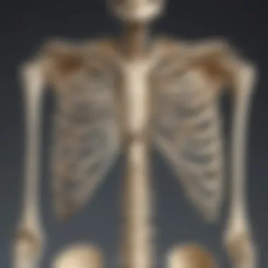
The rib cage protects the heart and lungs while providing support for the thoracic cavity. It consists of twelve pairs of ribs, along with the sternum.
- Key Characteristic: The rib cage's design allows for expansion and contraction, which is essential for respiration.
- Unique Feature: The ability of the ribs to move makes the rib cage adaptable to different states of breathing.
However, this adaptability can lead to delicate areas where fractures may occur if subjected to high impact.
Appendicular Skeleton
The appendicular skeleton includes the upper and lower limbs and the pelvis. This part of the skeleton is essential for movement and manipulation of objects.
Upper Limbs
The upper limbs include the shoulder girdle, arm, and hand bones. This skeleton configuration is designed for a wide array of motions.
- Key Characteristic: The flexibility of the shoulder joint allows for complex movements in different directions.
- Unique Feature: The presence of the clavicle connects the upper limb to the trunk, acting as a strut to stabilize the shoulder.


This flexibility is beneficial as it supports a variety of activities from lifting to throwing, though it is also more prone to dislocations than other joints in the body.
Lower Limbs
Lower limb bones provide support for the body’s weight and are integral for walking and running.
- Key Characteristic: The femur is the longest and strongest bone, supporting most of the body’s weight.
- Unique Feature: The knee joint allows for a greater range of motion, contributing to ambulation.
Yet, injuries can lead to significant mobility issues, especially if the femur is affected.
Pelvis
The pelvis connects the spine to the lower limbs and supports the organs of the lower abdomen.
- Key Characteristic: The pelvis plays a crucial role in body stability during movement.
- Unique Feature: Its shape provides a cradle for the reproductive organs.
However, its structure is complex, and abnormalities can lead to various health concerns.
The components of the human skeleton play pivotal roles in physical health and functionality. Understanding these components helps educators and caregivers foster knowledge about our body's design.
Detailed Anatomy of the Skull
The anatomy of the skull is a cornerstone in understanding the human skeleton. It serves significant functions such as protection of the brain, formation of the facial structure, and provision of attachment points for muscles. The skull is comprised of two main parts: the cranial bones, which encase the brain, and the facial bones, which shape our face. Understanding these elements is crucial for educators and caregivers as it helps illustrate the complexity of biological structures to children.
Cranial Bones
Cranial bones play an essential role in safeguarding the brain. They are formed by eight different bones, each contributing uniquely to both function and structure.
Frontal Bone
The frontal bone is the foremost cranial bone, forming the forehead and the upper part of the eye sockets. It is crucial for providing structural support to the face and protecting the frontal lobe of the brain. Its flat and robust nature makes it a popular focus in skull anatomy as it illustrates the importance of bone density in cranial protection. The frontal bone’s unique feature includes the frontal sinus, which helps in drainage and reduces the weight of the skull. In terms of disadvantages, a fracture in this bone can lead to serious injuries to the brain due to its position.
Parietal Bones
Parietal bones are paired structures that form the sides and roof of the skull. They connect with each other at the top of the head through the sagittal suture. Their primary role includes protection and giving shape to the cranial cavity. Parietal bones are significant due to their size and surface area for muscle attachment. A unique aspect of parietal bones is their varied thickness that can adapt to physical stress. However, their extensive nature means that damage can lead to severe implications, including intracranial injury.
Occipital Bone
The occipital bone forms the back of the skull and base of the cranium. It is vital for encasing the brain stem and provides an articulation point for the vertebral column. Key characteristics include the foramen magnum, which is an opening for the spinal cord. This bone is beneficial due to its supportive role in head movement. A unique feature is its occipital condyles, allowing the skull to rotate on the spine. On the downside, occipital fractures can impair basic functions such as vision or balance.
Temporal Bones
Temporal bones are located on either side of the skull, forming parts of the sidewalls and base. They house essential structures related to hearing and balance. Their importance lies in their contribution to the overall protection and function of the auditory system. Temporal bones are notable for containing the petrous part, which houses the inner ear. The disadvantage here includes the association with several vital nerves; an injury can result in hearing loss or facial weakness.
Facial Bones
Facial bones, consisting of 14 different bones, shape the face and provide structure for essential functions, such as eating and breathing. They contribute significantly to our identity and functionality.
Nasal Bones
Nasal bones are two small oblong bones forming the bridge of the nose. Their primary function is structural and protective, contributing to the shape of the nasal cavity. The nasal bones are significant as they are often the most frequently fractured facial bones. A unique feature is their connection to the maxilla, allowing for an integrated structure. However, their small size can lead to challenges in surgical interventions.
Maxilla
The maxilla is the upper jawbone, playing an essential role in holding the upper teeth and forming the base of the orbits. Its function is integral to chewing and speech. The maxilla is crucial for its broad structure, which supports various facial muscles. Unique to the maxilla is the maxillary sinus, providing airspace and reducing weight. Downsides include that fractures can severely impact both aesthetic and functional aspects of the face.
Zygomatic Bone
Zygomatic bones, or cheekbones, are prominent structures positioned below the eyes. They are significant to facial aesthetics and protect the eyes as well. Their notable characteristic is contributing to the lateral walls of the orbits, providing facial contour. One unique feature is their articulation with several other bones, which allows for facial flexibility. Generally, they are strong but can be prone to fracture due to their prominence.
Mandible
The mandible, or lower jawbone, is the largest and strongest facial bone. Its primary function is to hold the lower teeth and facilitate movement for chewing and speaking. Unique characteristics include its movable joint at the temporomandibular joint, enabling a range of motions. The advantage of the mandible is its robust structure, which withstands significant forces. Conversely, fractures in the mandible can lead to complex solutions for restoration and function.
Understanding the Vertebral Column
The vertebral column, often referred to as the spine, is a key component of the human skeleton. It plays a critical role in maintaining posture and supporting the body’s structure. This section will explore the different regions of the spine and the intervertebral discs, providing insights into their distinctive features and functions. By understanding the vertebral column, one appreciates how integral it is to overall health and movement.
Regions of the Spine
The vertebral column is divided into several regions, each serving specific functions and providing various levels of flexibility and strength. The spine consists of cervical, thoracic, lumbar, sacral, and coccygeal regions. Let us explore each region in detail.
Cervical Vertebrae
The cervical vertebrae are found in the neck region and consist of seven bones, labeled C1 through C7. The most notable characteristic of cervical vertebrae is their range of motion, which enables the head to turn and tilt in various directions. This mobility is vital for numerous everyday activities, such as looking up or around, and contributes to overall body stability.
A unique feature of cervical vertebrae is the presence of foramina, which are holes that allow arteries and nerves to pass through freely. This anatomical design provides advantage in protecting crucial blood vessels while also allowing enough space for nerve root passage. Issues in this area can lead to discomfort or restricted movement in the neck, impacting quality of life.
Thoracic Vertebrae
The thoracic vertebrae consist of twelve vertebrae labeled T1 through T12. They form the upper back and are connected to the rib cage, which stabilizes the chest. A key characteristic of thoracic vertebrae is their articulation with ribs, which allows for protection of internal organs, such as the heart and lungs.
One distinct feature of thoracic vertebrae is their limited range of motion in comparison to cervical vertebrae. This rigidity provides a solid support structure that is essential for protecting vital organs. However, the limitation in flexibility can be a disadvantage if excessive tightness occurs, leading to discomfort or pain.
Lumbar Vertebrae
The lumbar vertebrae are located in the lower back and consist of five vertebrae labeled L1 through L5. Prominent for their size, lumbar vertebrae are the largest in the entire vertebral column, designed to bear significant weight and support the upper body during various activities. Their key characteristic is the ability to withstand pressure from lifting and bending motions.
Their unique feature includes a wide spinous process which provides ample space for muscle attachment. This structure offers strong support and is particularly beneficial for mobility. However, disadvantages can arise from high stress on the lumbar area, which can lead to issues like lower back pain or herniated discs.
Sacrum and Coccyx
The sacrum and coccyx are located at the base of the vertebral column. The sacrum consists of five fused vertebrae that connect the spine to the pelvis. The coccyx, commonly known as the tailbone, is made up of three to five fused vertebrae. The key characteristic of the sacrum is its role in providing strength and stability to the pelvic region, which is crucial for supporting the weight of the upper body when standing or moving.
A unique feature of the sacrum is its triangular shape, which allows for a strong foundation. This region serves as an attachment point for various muscles and ligaments related to movement. Whereas the coccyx serves a beneficial function as it provides attachment points for muscles involved in movement and stability but can also be subject to injury or discomfort if pressure is applied excessively.
Intervertebral Discs
Intervertebral discs are fibrocartilaginous structures located between the vertebrae. They act as shock absorbers, allowing movement while preventing the stretching of spinal nerves. Composed of an outer annulus fibrosus and an inner nucleus pulposus, these discs have the essential function of providing cushioning and flexibility to the vertebral column.
"Understanding the structure of vertebral discs is pivotal for appreciating their role in spinal health and mobility."
In summary, the vertebral column serves as a critical support system for the body, enabling not only movement but also protection for the spinal cord. Understanding its structure aids in recognizing healthy practices to maintain spine health, especially in children who are still developing their spinal structures.
The Rib Cage: Structure and Function
The rib cage is an essential part of the human skeleton, playing a critical role in both protection and support. It encases vital organs such as the heart and lungs, thereby ensuring their safety from physical damage. Additionally, the rib cage contributes to the overall shape and structure of the human torso. Understanding the components and functions of the rib cage provides valuable insights into not only anatomy but also the importance of maintaining bone health.
Ribs Overview
The human rib cage consists of twelve pairs of ribs, each categorized into three types: true ribs, false ribs, and floating ribs. Each type has unique characteristics and functions that contribute to the rib cage's overall purpose.
True Ribs
True ribs are the first seven pairs of ribs that are directly connected to the sternum via costal cartilage. This direct attachment to the sternum is their most significant characteristic. True ribs play a crucial part in the rib cage structure, as they offer robust protection to the heart and lungs. Their solid connections support the thoracic cavity effectively. An advantage of true ribs is their stability, as they contribute significantly to the overall strength of the rib cage. Their position also allows for essential respiratory movements, aiding in the expansion and contraction of the lungs during breathing.
False Ribs
False ribs consist of the next three pairs, specifically the eighth, ninth, and tenth ribs. Unlike true ribs, false ribs do not have a direct connection to the sternum. They are attached to the seventh rib through shared costal cartilage. This indirect connection allows for greater flexibility in the rib cage. The key characteristic of false ribs is their ability to provide some protection while allowing for mobility and flexibility. This flexibility is advantageous during various movements, making the rib cage more adaptable to physical activities.
Floating Ribs


Floating ribs are the last two pairs of ribs. They are termed floating because they do not attach to the sternum or to the other ribs at all. Their uniqueness lies in their anatomical structure. Floating ribs provide limited protection as they are not anchored in place. Instead, they allow for greater movement of the lower back and abdomen. This increased mobility can be beneficial in certain activities, but it also means that floating ribs are more vulnerable to injury compared to true and false ribs.
Sternum
The sternum, or breastbone, is a flat bone located in the center of the chest. It serves as a fixture for the true ribs to attach, creating a stable structure for the rib cage. The sternum is divided into three parts: the manubrium, the body, and the xiphoid process. Each segment plays an integral role in maintaining the integrity of the rib cage.
The sternum not only supports the attachment of the true ribs but also protects the heart and major blood vessels. It is vital to overall respiratory function as it aids in the mechanics of breathing by expanding and contracting alongside the rib cage. Understanding the sternum's role highlights its importance in the anatomy of the skeletal system and its contribution to human health.
Bones of the Upper Limbs
The bones of the upper limbs play an essential role in allowing humans to perform a wide array of movements. This section focuses on the structure and significance of these bones, providing insights into how they function collectively to support daily activities. Understanding these compnents can help children appreciate the complexity of their own bodies, as well as the importance of maintaining bone health.
Shoulder Girdle
The shoulder girdle is crucial for upper limb functionality. It connects the arms to the trunk, allowing for a significant range of motion. This part of the skeleton consists of two major bones: the clavicle and the scapula, which work together to support arm movements.
Clavicle
The clavicle, commonly referred to as the collarbone, is a slender bone that runs horizontally across the upper chest. It serves as a bridge between the bones of the arm and the sternum, stabilizing the shoulder joint. One key characteristic is its S-shaped curve, which helps in distributing the weight of the arm during various activities. The clavicle also prevents shoulder dislocation by providing structural support.
A unique feature of the clavicle is its ability to withstand pressure. However, it can be susceptible to fractures, especially during sports activities. This makes it vital for physical education when teaching students about sports safety and anatomical awareness.
Scapula
The scapula, or shoulder blade, is a flat triangular bone situated at the back of the shoulder. It houses the glenoid cavity, which forms the socket for the arm bone to connect. This ability to articulate with the humerus enables a wide range of arm movements, making it a key player in both daily tasks and athletic performance.
One prominent characteristic of the scapula is its broad surface area. This is essential for muscle attachment, allowing for robust movement and coordination. While it is less likely to fracture than the clavicle, any injury to the scapula can lead to significant functional limitations. Knowledge of this bone's role is important for understanding shoulder mechanics in sports, particularly in activities like throwing or swimming.
Arm Bones
The bones of the arm include the humerus, radius, and ulna. Each of these bones contributes uniquely to arm structure and motion, facilitating diverse activities such as lifting, pushing, and gripping.
Humerus
The humerus is the longest bone in the upper limb, extending from the shoulder to the elbow. It is crucial for providing leverage and strength for the movements of the arm. One notable characteristic is the rounded head of the humerus, which fits into the shoulder socket, allowing for a great range of motion.
The humerus also contains various attachment sites for muscles, making it vital for actions such as lifting and throwing. Its length provides a mechanical advantage in many sports, highlighting its importance in both education and physical training.
Radius
The radius is one of the two long bones in the forearm, located on the thumb side. It plays a critical role in wrist movement and function. A key characteristic is its ability to rotate around the ulna, which enables supination and pronation—the motions of turning the palm up or down.
The radius is essential for activities that require wrist rotation, such as writing and throwing. Careful understanding of its structure can be beneficial for teaching proper hand techniques in sports and arts, emphasizing its role in daily life.
Ulna
The ulna, located on the opposite side of the radius, is slightly longer and forms the inner part of the forearm. Its primary role is to provide stability for the elbow joint. Notably, the ulna's trochlear notch articulates with the humerus to form a hinge joint, enabling flexion and extension of the arm.
One unique feature of the ulna is the olecranon process, which forms the point of the elbow. Understanding the ulna’s structure can help children learn about how their bodies bend and straighten during various movements, enhancing their comprehension of physical anatomy.
By learning about the bones of the upper limbs, children can better understand the importance of taking care of their bones through proper nutrition and exercise.
Bones of the Lower Limbs
The lower limbs are vital for movement, stability, and supporting body weight. This section focuses on two main groups of bones: the pelvic girdle and the leg bones. Understanding these components helps to appreciate their role in mobility and posture, particularly using the lower limbs during activities such as walking and running.
Pelvic Girdle
The pelvic girdle, a bony ring that connects the spine to the lower limbs, consists of three main bones: the ilium, ischium, and pubis. Each of these bones plays a crucial role in support and movement.
Ilium
The ilium is the largest bone of the pelvic girdle. It provides a broad surface for muscle attachment, and supports the weight of the body when sitting or standing. One key characteristic is its flared shape, which contributes to the overall structure of the pelvis. This design enhances stability, making it a popular choice for studies related to human movement and locomotion.
A unique feature of the ilium is the iliac crest, the uppermost part, which is easily felt on the side of the body. Its prominence can assist in anatomical education. However, its size can also make it prone to injuries, especially in sports.
Ischium
The ischium forms the lower and back part of the pelvis. It is vital for bearing weight when sitting, which is an important aspect of daily life. The ischium has a sturdy structure, providing strong support to the pelvic area. Its key characteristic is the ischial tuberosity, often referred to as the "sit bones." This specific structure allows us to feel comfort while sitting. The ischium can sometimes be less emphasized in skeletal studies, yet it is essential for understanding hip stability and lower limb function. Its depth allows for strong connections to ligaments and muscles.
Pubis
The pubis is the smallest bone in the pelvic girdle. It contributes to the front of the pelvis and helps to form the pubic symphysis. One of the significant aspects of the pubis is its role in sexual reproduction, as it houses structures important in childbirth.
A notable feature is that it meets with the other side at the pubic symphysis, allowing slight movement. This flexibility during movements can be beneficial, but can also lead to discomfort in some individuals.
Leg Bones
The leg bones include the femur, tibia, and fibula. Each of these bones contributes functionally to lower limb movement and weight-bearing activities.
Femur
The femur, also known as the thigh bone, is the longest bone in the human body. Its significant length is critical for mobility. The femur serves as the primary weight-bearing bone in the thigh area. One key characteristic is its round head that fits into the hip socket, facilitating a wide range of movements.
As a strong bone, the femur can withstand significant forces, making it a popular focus of studies in sports medicine. Its disadvantage is its length; fractures can be serious and may require surgical intervention.
Tibia
The tibia, commonly called the shinbone, is the larger and stronger of the two leg bones. It carries most of the weight in the lower leg. A key defining characteristic of the tibia is its flat upper surface, known as the tibial plateau, which connects to the femur at the knee joint. Being a weight-bearing bone makes the tibia an important focus in discussions related to injuries and rehabilitation. A disadvantage is its exposure to stress fractures, especially in athletes or individuals with high activity levels.
Fibula
The fibula is a slender bone that runs parallel to the tibia. It plays a supportive role rather than a weight-bearing one. The key feature of the fibula is its ability to provide stability to the ankle joint. This attribute is essential for proper walking and running mechanics. Although it is not a primary weight-bearing bone, it can still be affected by injury. Its minimal involvement in weight-bearing can be seen as an advantage, as recovery from fibula-related injuries is often quicker compared to the tibia or femur.
Joint Structures and Types
Understanding the types of joints and their structures is essential in the study of the human skeleton. Joints play a fundamental role in facilitating movement, providing stability, and allowing the skeletal system to function effectively. Proper knowledge of joint types aids educators and caregivers in explaining how our bodies move and adapt to various activities.
Types of Joints
Fibrous
Fibrous joints are characterized by their dense connective tissue, which limits movement. They primarily serve to provide stability. A key aspect of fibrous joints is that they are immovable. This type includes structures like sutures in the skull, where the bones are tightly bound together. The unique feature of fibrous joints is their ability to form strong connections between bones. These joints provide significant advantages in terms of protection for the brain and other vital internal structures. However, the lack of mobility can be seen as a disadvantage in certain scenarios.
Cartilaginous
Cartilaginous joints consist of cartilage that connects the bones. These joints allow for limited movement, making them more flexible than fibrous joints. A prominent example is the intervertebral discs in the spine. The key characteristic of cartilaginous joints is their ability to absorb shock, which is crucial for maintaining spinal health and overall mobility. This flexibility can be beneficial, especially in areas needing movement while still offering support. However, wear and tear can lead to issues that affect flexibility, presenting a disadvantage over time.
Synovial
Synovial joints are recognized for their wide range of motion. They are filled with synovial fluid, which lubricates the joints and reduces friction during movement. Examples include the knee and elbow joints. The key characteristic of synovial joints is their capability for extensive movements, which is vital for athletic activities and everyday actions. These joints can be classified into several types based on their movement, adding to their advantage. The disadvantage may include the susceptibility to injuries such as sprains or arthritis, affecting joint mobility and comfort.
Common Joint Movements
An understanding of common joint movements helps clarify how the skeleton functions during various activities. Movement is essential in daily life, whether it is walking, playing, or exercising.
Flexion and Extension
Flexion and extension are fundamental movements that involve bending and straightening a joint. Flexion decreases the angle between two body parts, while extension increases it. For instance, bending your elbow to bring your hand closer to your shoulder involves flexion. The key characteristic is that these movements are crucial for activities like walking and reaching. These movements are beneficial as they allow for greater range of motion. However, overexertion can lead to strains and injuries if not performed correctly.
Abduction and Adduction
Abduction refers to moving a limb away from the body's midline, whereas adduction means moving it toward the body. An example of abduction is lifting your arms sideways. The key characteristic of these movements is their vital role in exercises and physical activities. They are beneficial for maintaining balance and coordination. Yet, improper techniques can lead to muscle cramps or injuries, highlighting the importance of teaching correct methods.
Rotation
Rotation is the circular movement of a body part around its axis. This motion is seen in the shoulder and hip joints. The key characteristic is that rotation is important for activities such as turning or twisting. This movement offers significant benefits in various sports and exercises where changing directions is crucial. Nonetheless, excessive rotation can result in joint strain or injuries, especially in competitive settings.


"A solid understanding of joint structures fosters better coordination and physical activity, which is important for growing children."
Growth and Development of Bones
The growth and development of bones is a fundamental aspect of human anatomy. It plays a crucial role in overall health and physical capabilities. Understanding how bones grow is not only interesting but also important for children's health. Proper development of the skeleton ensures that children can move freely and engage in physical activities.
Bone Formation
Bone formation is the process by which new bone tissue is created. This is particularly important during childhood and adolescence when the body undergoes rapid changes. There are two main types of bone formation: intramembranous ossification and endochondral ossification.
- Intramembranous Ossification: This process primarily occurs in flat bones, such as the skull. In this type, bone directly forms from connective tissue, providing a framework for the new bone.
- Endochondral Ossification: Most bones in the human body grow through this mechanism. Here, cartilage serves as a template that is gradually replaced by bone. This method is significant for long bones like the femur and provides length and structure to the skeleton.
Bone formation is vital for developing strong and healthy bones. Nutritional factors, such as adequate calcium and Vitamin D intake, support this process. When children do not get enough nutrients, it can affect their bone growth and lead to potential health issues later in life.
Growth Plates
Growth plates, also known as epiphyseal plates, are regions of growing tissue located near the ends of long bones. They are crucial in determining the length and shape of bones as a child grows. Growth plates are made of cartilage and gradually turn into bone as a child ages.
- Location: The growth plates exist in various bones, especially long bones like the femur, tibia, and humerus.
- Significance: Growth plates are essential for any child's height. Once a child reaches adulthood, these plates close and the bones stop growing.
- Hormonal Influence: Growth hormones significantly influence the activity of growth plates. Factors such as puberty trigger increased hormone levels, leading to bone growth spurts.
Understanding how growth plates work is essential for recognizing potential growth disorders. Conditions affecting growth plates can lead to abnormal bone growth and height issues.
For further information, you can explore resources on Wikipedia and Britannica.
Engaging in activities that promote bone health at an early age can lead to a lifetime of benefits. Physical activity is equally crucial, as it encourages bone density and strength.
Importance of Bone Health
Bone health is a crucial topic that cannot be overlooked when discussing the human skeleton. Strong and healthy bones play a vital role in overall well-being and physical function. This section emphasizes why maintaining bone health is important, particularly for children aged three to twelve, who are in critical stages of growth.
Bone health contributes to effective movement, proper posture, and injury prevention. It is essential for supporting the body's structure as well as protecting internal organs. Healthy bones also store minerals such as calcium and phosphorus, which are necessary for various body functions. Moreover, they facilitate the production of blood cells within the bone marrow. Therefore, prioritizing bone health can lead to long-term benefits, including decreased risk of fractures and chronic conditions like osteoporosis.
Nutrition for Healthy Bones
A balanced diet rich in essential nutrients is vital for maintaining healthy bones. Two key components in this process are calcium and vitamin D, both of which play significant roles in bone development and maintenance.
Calcium
Calcium is an essential mineral, fundamental for bone structure and strength. It provides stability and density, which are necessary for supporting the body's weight. One key characteristic of calcium is its abundance; it is readily available in many food sources. Dairy products such as milk, cheese, and yogurt are often praised for their high calcium content. Additionally, leafy greens like kale and fortified products such as orange juice also contribute to adequate calcium intake.
A unique feature of calcium is its partnership with vitamin D. While calcium can be sourced from the diet, it needs vitamin D for optimal absorption in the body. Therefore, ensuring adequate calcium intake is particularly beneficial during childhood as it supports the developing bones' growth. However, excessive consumption without proper balance can lead to issues, highlighting the importance of moderation.
Vitamin
Vitamin D, often dubbed the "sunshine vitamin," is crucial for the absorption of calcium. It aids in making calcium available in the bloodstream, which is necessary for effective bone mineralization. This vitamin can be synthesized in the skin upon sunlight exposure, but dietary sources are equally important. Foods like fatty fish, egg yolks, and fortified cereals are good options for obtaining vitamin D.
A key characteristic of vitamin D is its unique ability to enhance the efficiency of calcium absorption. Thus, it is a popular choice in discussions about bone health. Moreover, maintaining sufficient levels of vitamin D supports immune function and overall health. Inconsiderate intake may pose risks, particularly during winter months or in areas with limited sunlight. Without adequate vitamin D, the benefits of calcium cannot be fully realized.
Physical Activity
Physical activity is another critical element for maintaining bone health. Engaging in regular exercise leads to stronger bones by stimulating bone remodeling. Activities such as running, jumping, and resistance training enhance bone density and overall strength.
Weight-bearing exercises are particularly effective as they encourage bones to work against gravity. Children should be encouraged to participate in various forms of physical activities to support their growing skeletons. Furthermore, physical activity helps improve balance and coordination, reducing the likelihood of falls and injuries.
Common Skeletal Disorders
In understanding the human skeleton, it is essential to recognize common skeletal disorders. These conditions can significantly affect overall health and mobility. By identifying these disorders, caregivers and educators can better support children who may be at risk or who are developing. Awareness of these issues can lead to early interventions, improving outcomes for affected individuals. Furthermore, knowledge of common disorders can help families understand the importance of maintaining bone health through proper nutrition and activity.
Osteoporosis
Osteoporosis is a condition that weakens bones, making them more prone to fractures. It is common in older adults but can also affect younger individuals, especially if they have poor diets or inactive lifestyles. Here are a few critical points about osteoporosis:
- Bone Density: Osteoporosis occurs when bone density decreases, leading to an increase in the fragility of the bones.
- Risk Factors: Factors like a lack of calcium and vitamin D in the diet, inactivity, and certain medications can contribute to osteoporosis.
- Symptoms: Often, there are no symptoms until a fracture occurs. Therefore, awareness and prevention measures are vital.
- Prevention: To prevent osteoporosis, it is essential to consume a balanced diet rich in calcium and spend time on weight-bearing exercises.
"Taking care of bone health early in life is key to preventing diseases like osteoporosis later on."
Scoliosis
Scoliosis is a lateral curvature of the spine that can occur during growth. It is crucial for parents and educators to recognize its signs. Here are the main points about scoliosis:
- Types: There are different types of scoliosis, including idiopathic, congenital, and neuromuscular. Idiopathic scoliosis is the most common and usually appears in puberty.
- Symptoms: Symptoms can include uneven shoulders or hips, and in severe cases, it may cause back pain or difficulty in breathing.
- Management: Treatment options depend on the severity of the curve. Mild cases may only require monitoring, whereas severe cases might need bracing or surgery.
- Importance of Early Detection: Early detection can lead to more effective treatment, preventing complications later in life.
Children, parents, and educators must have a basic understanding of these skeletal disorders. Knowledge allows for a proactive approach to bone health, ensuring children lead healthy lives.
Educational Activities and Resources
Understanding the human skeleton can be made more engaging through various educational activities and resources. These tools play a critical role in helping children grasp the complexities of the skeletal system. By providing interactive and practical experiences, they enhance the learning process. Additionally, these activities cater to different learning styles, making it easier for children to retain information.
Interactive games act as an excellent way to familiarize young learners with the bones and their functions. They encourage participants to think critically and apply knowledge in a fun context. Games focusing on anatomy can be especially helpful in reinforcing lessons. For example, digital skeleton labeling games allow children to click on different bones to learn their names and locations. This method promotes active engagement and retention.
DIY projects also offer hands-on experience, vital for understanding the skeletal structure. Creating models or skeletons can provide visual learners with a tangible representation of the human body. Activities such as building a skeleton from household materials such as cardboard or clay can be both educational and creative. Moreover, they encourage teamwork and communication, skills essential in any collaborative effort.
In developing these activities, it is important to consider the age and comprehension level of the participants. Tailoring resources to match their capabilities ensures better engagement. Educators and caregivers should aim to create a balance between challenge and accessibility to maintain interest without causing frustration.
By using these tools, we can deepen the appreciation and understanding of the human skeleton among the younger audience. They serve not only as teaching aids but also foster curiosity and exploration in biology.
"Engaging students with hands-on activities can lead to greater comprehension and interest in topics such as human anatomy."
Interactive Games
Interactive games are a dynamic approach to learning. These games can span digital platforms or physical activities. Online resources often incorporate quizzes, puzzles, and animation that visualize the skeleton's functions. In addition to digital games, role-play scenarios can bring a real-world aspect to the learning environment. For instance, creating a scavenger hunt where students find and label different bones in a classroom can cement their knowledge in an enjoyable way.
Some recommended online resources might include educational platforms like Britannica and discovery channels that host interactive tools for this purpose. These games typically feature incentives for successful completion, making learning both competitive and rewarding.
DIY Projects
DIY projects provide a tactile experience that reinforces theoretical learning. One idea could be to use pasta to create a skeleton model. By stringing different shapes of pasta together to represent bones, children can visualize and physically manipulate the parts of a human skeleton. This project makes the learning process not only informative but also enjoyable.
Another engaging project is making a poster-sized skeleton using craft materials. Students can research each bone's name and function and create labels to enhance their understanding.
Furthermore, these DIY projects foster creativity, allowing children to express their understanding of anatomical concepts in various forms.
In summary, educational activities focused on the human skeleton are vital in helping children understand this complex subject. They present information in a digestible manner and cater to various learning styles. Teachers, parents, and caregivers can leverage these resources for a richer learning experience.
End
The conclusion serves a vital role in understanding the comprehensive guide to the human skeleton. It encapsulates the key findings and insights that arise from the exploration of the skeletal system. This section is not merely a summary, but it reinforces the relevance of the topic by emphasizing its importance in education and health.
One specific element is the recap of key points related to the structure and function of bones. A well-structured conclusion brings together various aspects covered in earlier sections. This might involve reiterating how bones offer support, enable movement, protect vital organs, store minerals, and produce blood cells. Such a reminder is crucial for reinforcing knowledge, especially for children who are the primary audience.
Another benefit of this section is the opportunity to reflect on the broader significance of bone health. Understanding how nutrition and physical activity contribute to strong bones sets the stage for lifelong habits. Parents, teachers, and caregivers can instill the importance of bone health from a young age. This knowledge can lead to informed decisions regarding diet and exercise, promoting overall well-being.
Thus, the conclusion does more than round off the article. It provides a thoughtful reflection on how the human skeleton's structure and functions play critical roles in everyday life. The insights gleaned in this guide empower readers to appreciate their bodies and take proactive steps toward maintaining bone health.
Recap of Key Points
To summarize the core messages of the guide:
- The human skeleton consists of two main parts: the axial and appendicular skeletons.
- Bones are classified into cortical and trabecular types, each serving unique functions.
- The skeleton supports the body, facilitates movement, safeguards organs, stores minerals, and produces blood cells.
- Healthy bones rely on adequate nutrition, particularly calcium and vitamin D, as well as regular physical activity.
In addition, common disorders like osteoporosis and scoliosis highlight the necessity of bone health awareness.
Future Learning Opportunities
To extend knowledge beyond this guide, there are several avenues for further exploration:
- Interactive learning platforms, such as educational games or apps, can engage children and deepen their understanding of skeletal anatomy and functions.
- DIY projects that involve building models of the skeleton can serve as practical applications of the concepts learned.
- Websites like Wikipedia and Britannica provide extensive resources that are continuously updated, making them excellent sources for further research.
- Discussions on platforms like Reddit can provide insights and community support for educators and learners alike.
By encouraging further exploration, readers can obtain a holistic view of the human skeleton and its vital role in both health and education.


